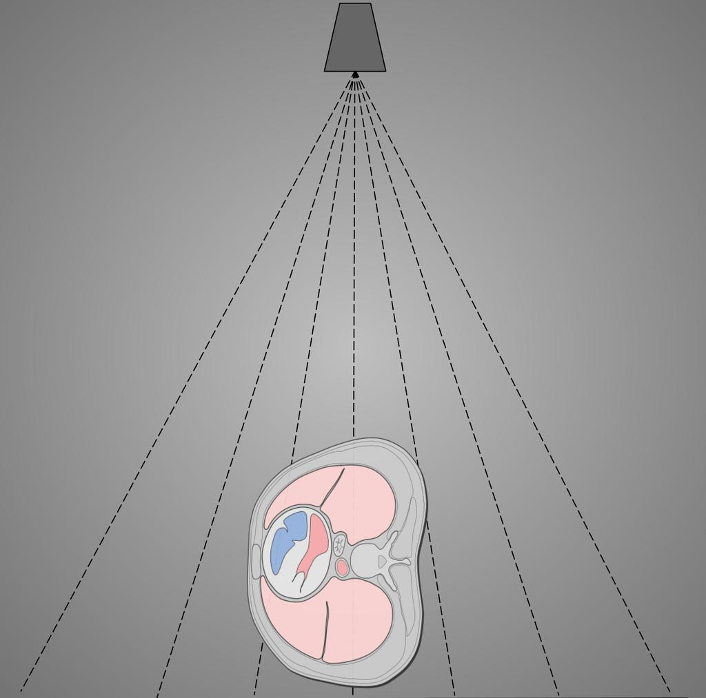
Using the paravertebral gutter technique see figure 1 right side rotated 5 10 anterior.
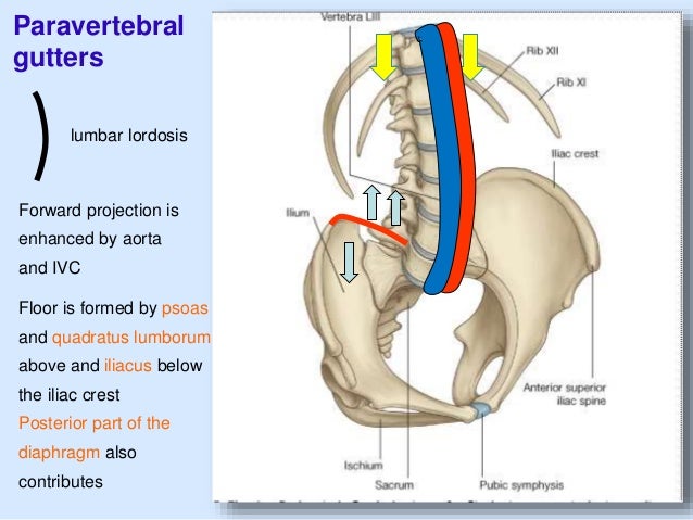
Left paravertebral gutter.
Patients present with signs and symptoms of pulmonary infection of a lower lobe mass.
An enlarged left auricle is frequently found in combination with dilated pulmonary artery branches and prominent pulmonary conus.
It joins the iliacus muscle to form the iliopsoas muscle the strongest hip flexor of the human body.
The midcoronal plane of the level of the 7 th thoracic vertebra approximately the inferior angle of the scapulae collimation.
Psoas major is a triangular bilaterally paired muscle that forms part of the floor of the paravertebral gutter.
The deep vertical recess formed on either side of the thoracic cage by the posterior curvature of the ribs and containing the posterior portions of the lung.
Sulcus pulmonalis ta paravertebral gutter pulmonary sulcus farlex partner medical dictionary farlex 2012 want to thank tfd for its existence.
We would like to show you a description here but the site won t allow us.
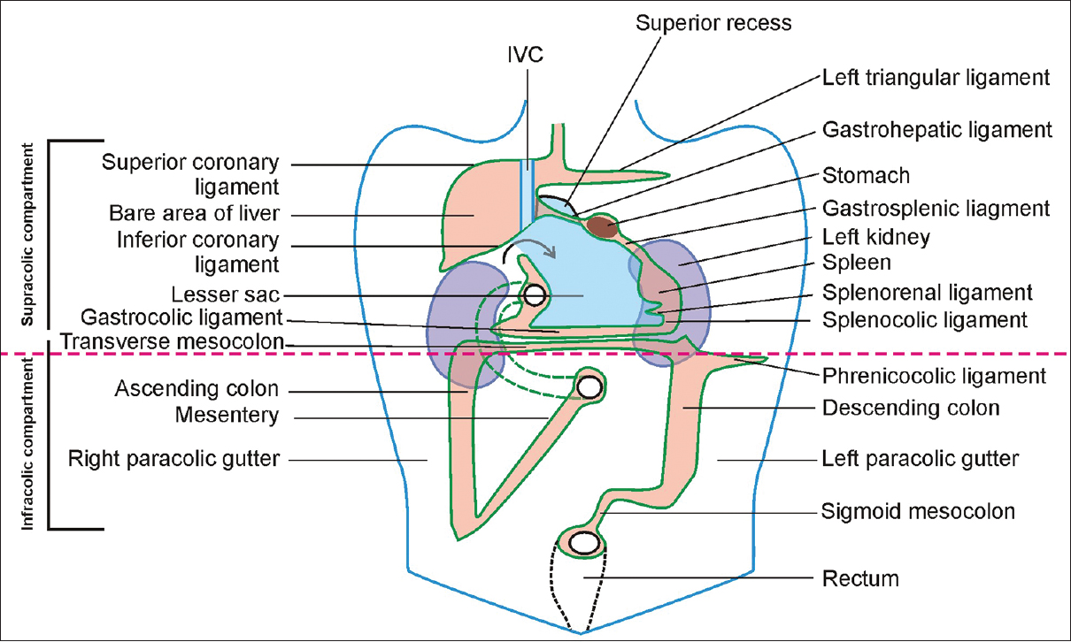
The left medial paracolic gutter.
1 article features images from this case.
The left auricle is the first chamber to enlarge in mitral stenosis and may reach an enormous size owing to hypertrophy and dilatation.
The main paracolic gutter lies lateral to the colon on each side.
In addition the lordotic lumbar spine has the right and left paravertebral gutters on either side.
In most cases of long standing x ray findings are produced which are diagnostic.
Chest lateral view 1 public playlist include this case.
Iliopsoas is important for standing walking and running.
The right and left paracolic gutters are peritoneal recesses on the posterior abdominal wall lying alongside the ascending and descending colon.
On the patient s scapulae and place the patient into rao until the light from the light beam diaphragm is seen on the left thumb.
The anterior pole of the spleen lies immediately supero lateral to the splenic flexure of the colon see figure 1 and just above the phrenicocolic ligament.
Unlike extralobar sequestration it is rarely associated with other developmental abnormalities.
The posterior pole of the spleen lies in or just lateral to the left paravertebral gutter level with the first lumbar vertebra.
There is a ventral curvature or lordosis of the lumbar vertebrae which is enhanced by the inferior vena cava and the aorta.
Paravertebral gutter technique diagram.