Protrusio acetabuli and acetabular retroversion a posteriorly oriented acetabulum can be seen on oblique axial mri of the hip.
Left acetabular roof mri.
Radiotherapy effectively decreases pain yet it does not restore the ability to load the joint.
It is seen in as many as 10 of hips and is typically located at the 12 o clock position both in the coronal and sagittal planes.
Type 1 which was filled with contrast material on mr arthrograms and type 2 which was filled with cartilage.
22 acetabular depth can be quantified on the same oblique axial images that are used to calculate the alpha angle.
Epidemiology acetabular fractures are uncommon.
The reported incidence is approximately 3 per 100 0.
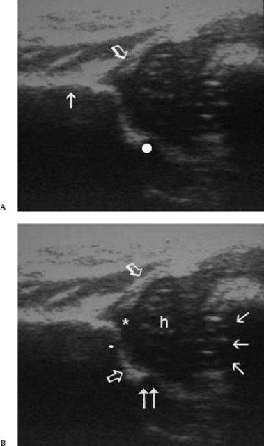
Subsequent mri was performed and demonstrated bone marrow oedema bmo in the acetabulum and the femoral head and neck figure 2.
The acetabular retroversion will result in an increase in coverage of the anterior aspect of the femoral head.
It is located more medially within the acetabular roof than the saf immediately adjacent to the acetabular notch figure 5.
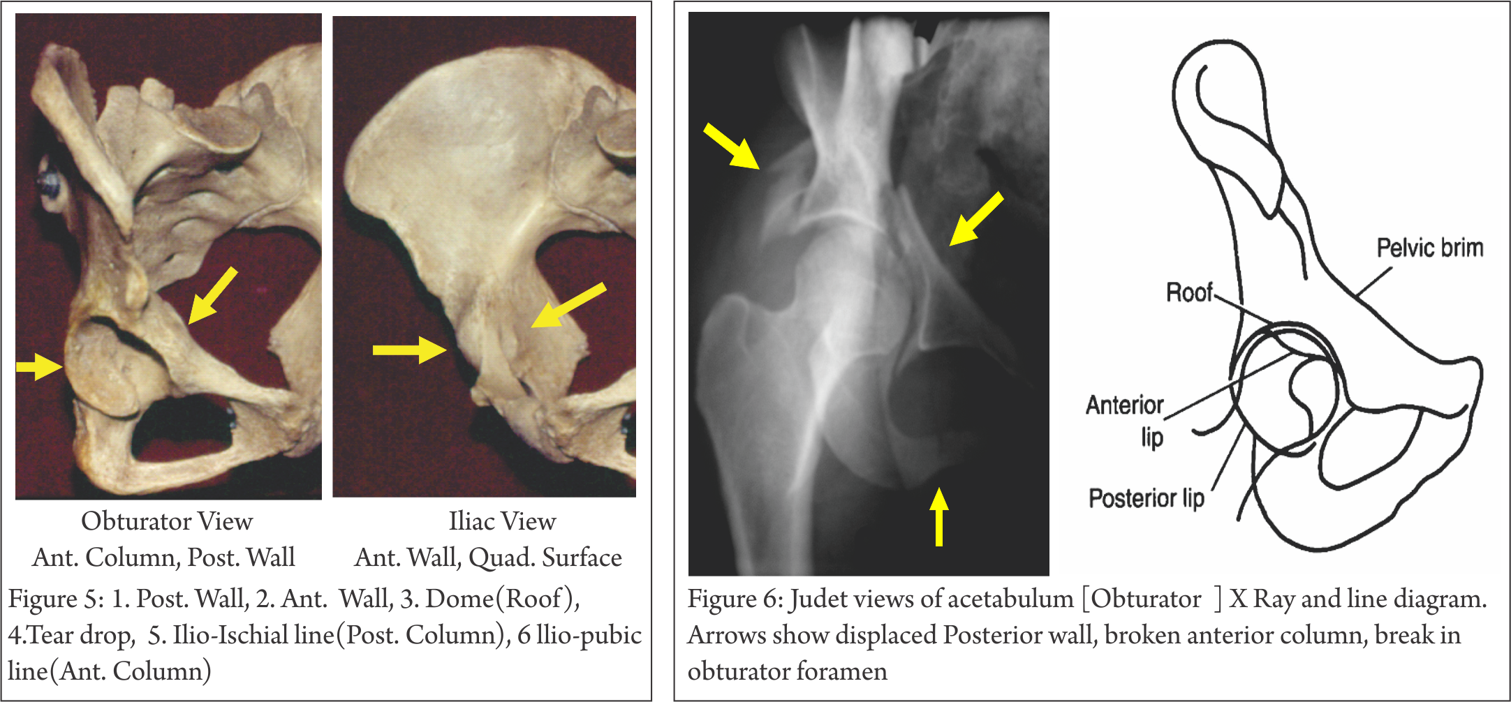
Acetabular fractures are a type of pelvic fracture which may also involve the ilium ischium and or pubis depending on fracture configuration.
A supra acetabular fossa also known as pseudodefect of acetabular cartilage is an anatomic variant whereby a focal defect is evident within the subchondral bone of the acetabular roof.
Often the initial radiographs are normal especially in the elderly os teoporotic patient.
Figure 1 bone scintigraphy anterior view showed intense uptake in the left acetabular region and less avid uptake in the femoral head and neck compared with the right side.
Saf was classified into two types.
Three dimensional surface rendered ct image of the right acetabulum obtained in anatomic position with digital transection in the coronal plane shows a 45 roof arc angle and a 10 mm subchondral arc purple area the superiormost 10 mm of the acetabulum and the arc subtended by the 45 roof arc angle.
Surgical treatment involving resection of metastatic lesions and joint reconstruction using bone grafts is burdened with a high rate of complications.
Metastatic lesions localized in the periacetabular area cause troublesome pain and reduced mobility of the patients.
A medical student a radiology fellow and two senior radiologists reviewed 1002 consecutive mr arthrograms for the presence of an accessory bony fossa in the roof of the acetabulum or saf.
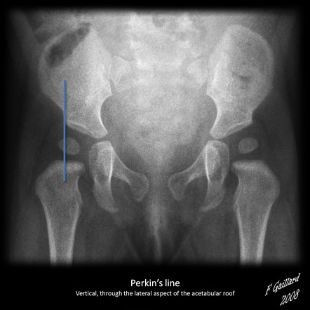
A coronal fat saturated t2 weighted mr image of pelvis shows deficient acetabular roof in left hip center edge angle 20 consistent with hip dysplasia.
In the athlete with a suspected stress fracture it is best to choose mri as the first advanced imaging test.